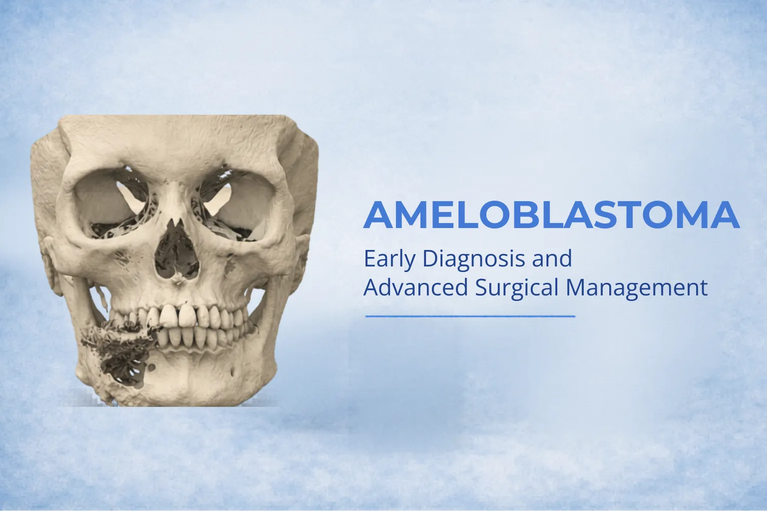
Ameloblastoma is a benign but locally aggressive odontogenic tumor arising from the enamel-forming cells of the jaw. Despite being non-cancerous, it has a high recurrence potential if not treated adequately. Early diagnosis and definitive surgical management are critical to prevent extensive jaw destruction and functional impairment.
What is Ameloblastoma?
Ameloblastoma typically originates in the mandible (lower jaw), especially in the molar–ramus region. It grows slowly but can expand significantly, causing facial asymmetry and bone destruction.
Key Characteristics:
- Benign but aggressive
- Slow-growing, often painless
- High recurrence if inadequately treated
- Can cause cortical expansion and tooth displacement
Causes and Risk Factors
The exact cause remains unclear, but it is believed to arise from:
- Remnants of dental laminaR
- Reduced enamel epithelium
- Odontogenic cyst lining
There are no well-established lifestyle risk factors, but it is commonly seen in: - Adults between 20–50 years
- Slight male predilection (variable)
Signs and Symptoms
Patients often present late due to the painless nature of the lesion.
Common Symptoms:
- Jaw swelling (most common)
- Facial asymmetry
- Loosening or displacement of teeth
- Malocclusion
- Occasionally pain or paresthesia
Types of Ameloblastoma
1. Conventional (Solid/Multicystic)
- Most common and aggressive
- High recurrence rate
2. Unicystic Ameloblastoma
- Often resembles a cyst
- Less aggressive
- Better prognosis
3. Peripheral Ameloblastoma
- Occurs in soft tissue (gingiva)
- Rare and less invasive
4. Desmoplastic Variant
- Mixed radiographic appearance
- Can mimic fibro-osseous lesions
Diagnosis
Accurate diagnosis requires a combination of clinical, radiographic, and histopathological evaluation.
Investigations:
- OPG (Orthopantomogram) – “soap bubble” or multilocular radiolucency
- CT Scan / CBCT – extent and cortical involvement
- Biopsy – definitive diagnosis
Treatment Options
1. Conservative Treatment
- Enucleation and curettage
- Indicated in select unicystic cases
- Higher recurrence risk
2. Radical Surgical Resection (Gold Standard)
- Marginal or segmental mandibulectomy
- Adequate safety margins reduce recurrence
- Often combined with reconstruction
3. Reconstruction Options
- Reconstruction plates
- Bone grafts
- Microvascular free flap (Fibula flap) – gold standard for large defects
- 3D planning and patient-specific implants
Why Early Treatment is Crucial
Delayed treatment can lead to:
- Extensive bone destruction
- Facial deformity
- Complex reconstruction
- Increased recurrence risk
Early intervention allows simpler surgery and better outcomes.
Prognosis and Follow-Up
- Recurrence rate depends on treatment modality
- Higher in conservative approaches
- Long-term follow-up (5–10 years) is essential
Expert Care in Mumbai
At our clinic in Mumbai, we provide:
- Advanced imaging and diagnosis
- Evidence-based surgical management
- 3D planning and reconstruction
- Personalized patient care
With expertise in maxillofacial tumor surgery and reconstruction, we ensure optimal functional and aesthetic outcomes.
When Should You Consult a Specialist?
Seek evaluation if you notice:
- Persistent jaw swelling
- Facial asymmetry
- Unexplained tooth mobility
- Non-healing oral lesions
Early consultation with a Maxillofacial Surgeon can significantly improve prognosis.
Conclusion
Ameloblastoma, though benign, demands aggressive and well-planned management. With modern surgical techniques and reconstructive options, patients can achieve excellent functional and aesthetic recovery.
Timely diagnosis and expert care remain the cornerstone of successful treatment.
Book a Consultation
If you or your patient is suspected to have ameloblastoma, consult:
Dr. Abhishek Chauhan
Maxillofacial & Oral Cancer Surgeon, Mumbai